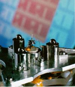
A scanning tunelling microscope filmed during operation by an electron microscope
A scanning electron microscope (SEM) is used to film the scanning tunneling microscope during operation. The principle of operation of an STM is visualized in this movie. A sharp tip is scanned over the surface and an image of the surface morphology is built up line by line like in the TV.
The STM-measurement is illustrated in the SEM-movie: It starts with a low magnification overview of sample and STM-tip. The sample is a ruthenium crystal covered with small Pb-particles. Increasing the magnification makes the lead particles become well visible with the tip shadow indicating that the tip is in tunneling contact with one of them. After the zoom the tip starts scanning this particle with a scan range of 5 µm
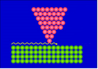
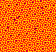
If the tip is so sharp that only a single atom is at its end, single atoms at the surface can be observed.
It is amazing that such high resolution can be achieved by an STM instrument with the size of a matchbox
In this video, a microscope probing on a nanoscale is filmed with another microscope during operation. We invite you to take a journey with us through the nanoworld and ultimately explore atomic structures. All movies shown here are made from original microscopy data.