Revealing whole-brain causality networks during guided visual searching
17th February 2022
Christian M. Kiefer, Junji Ito, Ralph Weidner, Frank Boers, N. Jon Shah, Sonja Grün, and Jürgen Dammers
In order for us to effectively interact with the environment in our daily lives, we use eye movements to actively sample the visual information around us. This is known as active vision. Given the vast amount of information seen at any one time, is important for the brain to process information selectively in line with contextual priorities.
Although previous studies have investigated task-dependent selective information processing, the way in which goal-directed processing of naturalistic visual stimuli affects the functional networks in the brain during active vision remains unclear.
Typically, studies focussing on visual processing in the human brain have avoided the use of high complexity of naturalistic stimuli in favour of artificial and simple stimuli, such as bars, gratings, letters, or simplified scenarios like controlled saccade tasks. However, while artificial stimuli make it easier to reduce the complexity of studies, responses to artificially simplified stimuli may not reflect neural responses to natural scenes.
Here, in a first of its kind study, magnetoencephalography was combined with eye-tracking technology to investigate how interregional interactions in the brain change when engaged in two distinct forms of active vision: freely viewing natural images or performing a guided visual search. Regions of interest with significant fixation-related evoked activity were identified with spatiotemporal cluster permutation testing.
The figure below shows causal interactions between brain regions in the alpha band in response to fixations, which were significant during free viewing (red) or visual searching (blue). Grey arrows indicate comparable connection strengths for both tasks.
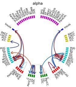
Using generalised partial directed coherence, the study shows that, in response to fixation onset, a bilateral cluster consisting of four regions (posterior insula, transverse temporal gyrus, superior temporal gyrus, and supramarginal gyrus) formed a highly-connected network during free viewing. Furthermore, a comparable network also emerged in the right hemisphere during the search task, with the right supramarginal gyrus acting as a central node for information exchange.
Based these findings, the researchers hypothesise that, following a fixation, the right supramarginal gyrus supplies the right supplementary eye field with new information to update the priority map guiding the eye movements during the search task.
As vision is, generally, the brain’s primary way of accessing information about the world, developing our understanding of the mechanisms involved in visual processing is important for a range of fundamental applications involving cognitive interactions. Future research will now also focus on a visual memory task.
Original publication:
Revealing whole-brain causality networks during guided visual searching