
TECHNICAL ASPECTS OF POSITRON EMISSION TOMOGRAPHY
Typically, a PET-scanner consists of a detector ring with 60 cm diameter and 20 cm length inside which the patient is positioned. For brain studies, the patient bed is positioned in such a way that the entire brain is within the field of view, whereas for full-body investigations, the bed is moved into the field of view step-by-step.
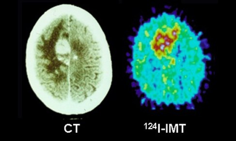
The detector consists of scintillation detectors with scintillation crystals coupled to photomultipliers. The radiation, which is emitted from the patient after the injection of a weak radioactively labelled substance (radiotracer), causes light pulses within the scintillation crystals. This light is then converted to electrical signals by the photomultiplier. A detector is linked to opposing detectors by a coincidence logic: if the radiation hits two opposing detectors within a time interval of, e.g. 6 ns, it is assumed that the radiation comes from a radiotracer molecule located on the line connecting the two detectors. During the PET study, which can last from a few minutes up to two or three hours, thousands of such coincidence lines are recorded. Using this data and appropriate programs, images are reconstructed which show the spatial and temporal distribution of the radiotracer within the patient. In this way, increased or decreased uptake in a tumour or an infarct area, respectively, can be visualised. Today’s PET scanners offer an image resolution of 4-5 mm.

Group Leader

- Institute of Neurosciences and Medicine (INM)
- Medical Imaging Physics (INM-4)
Building 15.2v /
Room 310
Room 310
+49 2461/61-96524
E-Mail
Staff

Dr.rer.medic. Cláudia Régio BrambillaPostdoctoral researcherBuilding 15.2v / Room 308+49 2461/61-5738
Last Modified: 09.03.2023