
PET
Positron emission tomography (PET) is a unique modality for imaging normal and diseased metabolical functions in vivo, in a quantitative manner.
To allow for optimum image quality, it is necessary to continuously improve the physical, technological and methodological prerequisites of simultaneous recording. For this purpose, we work to develop innovative reconstruction programs and procedures to correct for the different influences on the quantitative accuracy of PET. Furthermore, methods to extract and analyse diagnostically relevant parameters are studied.
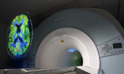
Although PET is the most sensitive tool for metabolic in vivo imaging, magnetic resonance imaging (MRI) delivers unsurpassed anatomical images and additional functional information, which is complementary to that obtained from PET. The hybrid 3T MR-PET and 9.4T MR-PET scanners installed in our institute open new frontiers by utilising both imaging modalities simultaneously. This advance in technology means that new physical and methodological challenges must be addressed. Important issues relate to the assessment of the new PET detector technology, corrections for attenuation, scatter and motion correction, as well as PET reconstruction guided by MRI.
The hybrid scanners enable the simultaneous highlighting of activated brain networks via fMRI and a representation of the biochemistry using PET. Furthermore, the hybrid MR-PET scanner is also able to serve as a platform to validate MR-based measurements of biochemical and physiological functions with the help of PET.
WORKING GROUPS
TECHNICAL ASPECTS OF POSITRON-EMISSION-TOMOGRAPHY
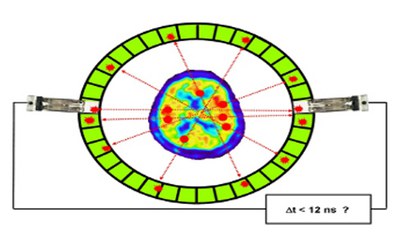
Typically, a PET-scanner consists of a detector ring with 60 cm diameter and 20 cm length inside which the patient is positioned. For brain studies the patient bed is positioned in such a way that the entire brain is within the field of view.
Technical Aspects of MR-PET
The hybrid scanner 3TMR-PET consists of a high resolution BrainPET, newly constructed by Siemens, and a commercial 3T MAGNETOM Trio MRI scanner.
Group Leader
Staff


Dr.rer.medic. Cláudia Régio BrambillaPostdoctoral researcherBuilding 15.2v / Room 308+49 2461/61-5738

Last Modified: 14.02.2023