Development and optimisation of MR-PET applications
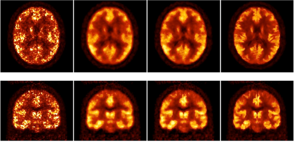
Dynamic PET studies measure activity distribution over time. The PET scan acquires data with higher temporal precision (list-mode acquisition), which can be later framed. That is to say, data acquired in a certain time interval is grouped together and reconstructed (Figure 1). Short time frames are necessary to monitor rapid changes in activity. For obtaining the physiological parameters, it is necessary to apply kinetic models to the activity distribution over time [1].
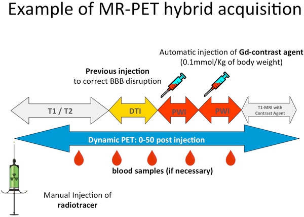
Examples of dynamic studies are those performed on brain tumour patients after injection of [18F]-Fluor-Ethyl-Tyrosine (FET), which lasts for 50 minutes. During the PET acquisition, a number of MR sequences can be performed. For example, T1-weighted sequences (with and without contrast medium), T2-, UTE-, and fMRI sequences, imaging related to blood flow such as PWI or ASL, and chemical shift imaging (CSI) can all be acquired simultaneously to the PET acquisition. However, the related sequences must match the temporal and spatial conditions of the PET protocol study as well as the aim of the study. For this purpose, MR protocols commonly used in MR-only studies must be adapted appropriately with respect to image volume, voxel size, signal-to-noise ratio, and acquisition time.
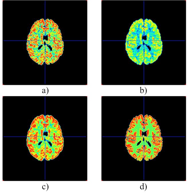
Simultaneous MR-PET data acquisition allows for dynamic studies in both modalities at the same time (Figure 2). The goal of these dynamic studies is to provide information relating to physiological parameters. These parameters can be represented in parametric brain images in a voxel-wise manner to preserve spatial resolution. The evaluation of different parametric brain images brings new understanding of the brain and its diseases. This is a new field of medical imaging called Parametric Imaging. For PET imaging, these parameters can include glucose concentration (Figure 3), binding potential for neuroreceptors, and blood flow, amongst others. For MRI, in addition to the structural images, different parametric maps can also be obtained such as, cerebral blood flow (CBF) or permeability (parameter used for brain tumours). In MRI, it is possible to generate parametric maps with techniques such as arterial spin labelling (ASL) (link to MR/PET ASL page), dynamic contrast enhanced (DCE) MRI, and dynamic susceptibility contrast (DSC) MRI.
- L. Caldeira, J. Scheins, P. Almeida, & H. Herzog, (2013) Evaluation of two methods for using MR information in PET reconstruction. Nuclear Instruments and Methods in Physics Research Section A: Accelerators, Spectrometers, Detectors and Associated Equipment, 702, 141-143.
- L.L. Caldeira, N. da Silva, J. Scheins, M.E. Gaens, & N.J. Shah (2015). Effects of regularisation priors and anatomical partial volume correction on dynamic PET data. IEEE Transactions on Nuclear Science,62(4), 1725-1731.