Attenuation Correction for MR-PET
The combination of Positron Emission Tomography (PET) with magnetic resonance imaging (MRI) in hybrid MR-PET scanners offers a number of advantages when investigating brain structure and function. A critical step of PET data reconstruction is attenuation correction (AC). Unfortunately, present hybrid MR-PET scanners do not offer the possibility for a PET-based measured attenuation correction. However, to achieve qualitatively and quantitatively acceptable PET images, data on tissue absorption must be available as a prerequisite of attenuation correction. A straightforward approach to solving this problem is to derive the attenuation data from MR images, which are typically recorded in MR-PET scanners, for anatomical information. Unfortunately, in contrast to the CT information in a PET/CT scanner, there is no relationship between the MR information and the attenuation coefficients needed for PET attenuation correction. This means that alternative methods are required. In order to address this, our institute has developed the template-based attenuation (TBA) method [1], and a method based on an artificial neuronal network (ANN) is currently under development [2]. The TBA method uses templates of attenuation maps which have been obtained from conventional PET-based attenuation images. This method is, at present, applied in routine MR-PET measurements, and although this approach works quite satisfactorily in general, it may fail if the patient shows extremely unusual or abnormal bone structures, e.g. after surgery. Therefore, further investigations of the use of ultrashort MR sequences (UTE) are being undertaken. The advantage of such MR images is the recognition of bone, which is not visible in the anatomical images.
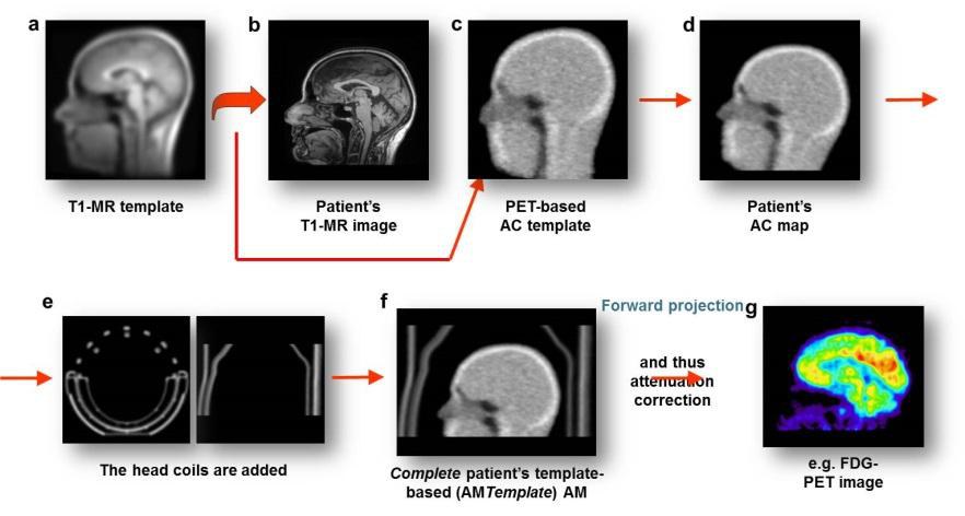
Template-based AC Method [1]
ANN-based AC Method [2]
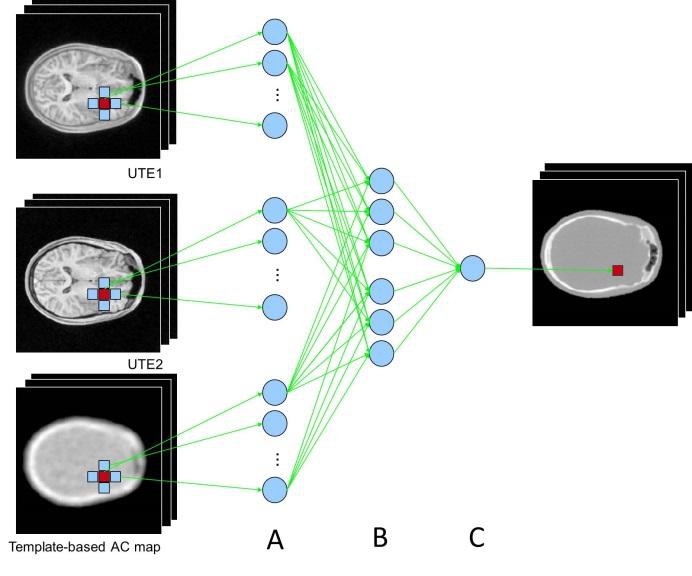
The UTE1 and UTE2 images are produced as a result of the double echo DUTE sequence. Note, the still visible signals from bone tissue in the UTE1 image acquired at ultrashort-echo time 0.07 ms.
Illustration of the methodology for attenuation correction of PET data with the proposed FFNN algorithms. The input features (A) are given to the FFNN (B) for the calculation of the attenuation coefficient (C). The generated attenuation images are then directly used for attenuation correction of PET data.
- E. Rota Kops, H. Hautzel, H. Herzog, G. Antoch, N.J. Shah (2015) Comparison of template-based versus CT-based attenuation correction for hybrid MR/PET scanners, IEEE Trans Nucl Sci, 62(5), 2115-2121
- A. Santos Ribeiro, E. Rota Kops, H. Herzog, P. Almeida (2014) Hybrid approach for attenuation correction in PET/MR scanners, Nucl Instrum Methods Phys Res, A734, 166–170