Not superimposable
Microscopists detect quasi-chiral walls in ferroelectric materials
2 September 2016
The atomic structure of ferroelectric materials is more diverse than previously assumed. An international research team made this discovery through experiments with electron microscopes carried out at the Ernst Ruska-Centre (ER-C) in Jülich. The ER-C is a leading centre for ultra-high-resolution electron microscopy. Their discovery could open up new fields of applications for a particular class of polarised crystals: so-called ferroelectric materials. The researchers believe these applications could range from the development of miniaturized pressure sensors and the tiniest of transistors through to highly integrated storage media (Nature Communications, DOI: 10.1038/ncomms12385).
Ferroelectric materials are today used for example in atomic force microscopes, where they enable the precise scanning of the tiniest surface details, even of individual atoms. Characteristic for this class of materials is an uneven distribution of charges, known as “electric polarization”. The reason for this is that ions are displaced slightly from their originally designated positions. The direction of the polarisation can be reversed by applying a voltage. However, the polarisation is not uniform throughout the material; small areas of varying polarisation, known as “domains”, are spread throughout it in a patchwork manner. These are separated by non-polarised layers that have thicknesses of just a few atoms, so-called “walls”.
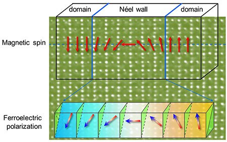
Researchers from Jülich, Lausanne in Switzerland, Xian in China and Burnaby in Canada, have now found that there are ferroelectric walls which are quasi-chiral. Their crystal structures cannot be superimposed, but behave like mirror images of each other, just like a person’s left and right hands. “Quasi-chirality distinguishes these walls from all others that were previously known”, explained Dr. Xiankui Wei from the Swiss research institute in EPFL, who is currently working as a research scientist in the Peter Grünberg Insitute in Jülich.
“This discovery may open up a new avenue towards designing novel nanoelectronic devices”, says Wei. “Theoretical studies have indicated that polarization rotation in ferroelectric lattices may trigger a giant electromechanical response. This effect could one day be used in miniaturized electronic components, such as tiny pressure sensors. Research on ferroelectric materials is still at an early stage, but it will be given a new boost by our discovery as we need to gain a more accurate understanding of how the formation of the walls can be controlled.”
The existence of chiral walls in ferromagnets is already known; there, they separate differently oriented magnetic areas. Detecting ferroelectric walls is however a much more difficult task, as these are usually thinner and the adjacent domains very small, measuring a maximum of 20 nanometres in size.
The ER-C in Jülich has now managed to achieve something of a scientific tour de force. “The ultra-high-resolution electron microscopes at the ER-C, which are among the best in the world, have made it possible to measure displacements of just a few picometres in the ions of our crystal samples, enabling direct imaging of chirality at the domain walls”, clarified Wei. One picometre is equal to a billionth of a millimeter. Moreover, a method developed in Jülich to strengthen the contrast of electron microscope images was employed to carry out this type of direct imaging and observation.
Original publication:
Néel-like domain walls in ferroelectric Pb(Zr,Ti)O3 single crystals;
Xiankui Wie et al.;
Nature Communications 7, published 19 August 2016, DOI: 10.1038/ncomms12385
Further information:
Peter Grünberg Institute – Division “Microstructure Research” (ER-C-1/ PGI-5)
Ernst Ruska-Centre for Microscopy and Spectroscopy with Electrons (ER-C)
Contact:
Dr. Xiankui Wei, Ernst Ruska-Centre for Microscopy and Spectroscopy with Electrons and Peter Grünberg Institute – Division “Microstructure Research” (PGI-5),
Forschungszentrum Jülich, Tel. 02461 61-9338, email: x.wei@fz-juelich.de
Press contact:
Angela Wenzik, Science Journalist, Forschungszentrum Jülich,
Tel. 02461 61-6048, email: a.wenzik@fz-juelich.de