A robust method for detecting small changes in the vicinity of the substantia nigra in Parkinson’s disease patients
24th February 2021
Krzysztof Dzieciol, Elene Iordanishvili, Zaheer Abbas, Adjmal Nahimi, Michael Winterdahl, N. Jon Shah
Parkinson’s disease is a progressive neurodegenerative disorder associated with involuntary shaking, stiffness of muscles and slow movement. Parkinson’s disease is caused by a loss of nerve cells in a part of the brain called the substantia nigra, which leads to a reduction in dopamine in the brain.
Consequently, in order to understand more about Parkinson’s disease, further detailed research related to the substantia nigra is required. Previous MR studies have attempted to achieve this using quantitative MR parameters based on delineated image provided by one of the employed sequences. However, this approach introduces a source of bias, as each sequence is sensitive to different biological changes in the tissue and hence the shape and size of pathological region might differ
To combat this problem, scientists from the INM4 have proposed a new method to investigate and reveal changes in quantitative MRI parameters in the vicinity of substantia nigra without any a priori delineation.
The approach uses an alternative method of statistical, voxel-based analysis of quantitative maps and was tested on 18 patients and 15 healthy controls using a well-established, quantitative free water mapping protocol.
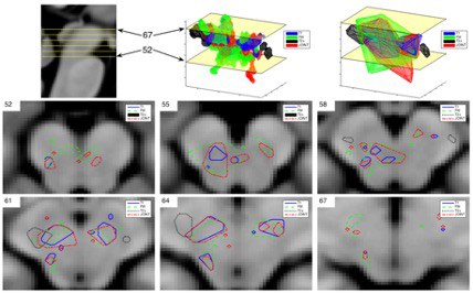
The results show that by applying the joint metric, involving three quantitative parameters (T1, T2*, free water content), it was possible to reveal the topology and the location of pathological changes in the substantia nigra and its vicinity. Moreover, a decrease in free water content, T1 and T2* in the vicinity of substantia nigra was indicated in the Parkinson’s disease patients compared to the healthy controls. These findings reflect a disruption of grey matter and iron accumulation, which is known to lead to neurodegeneration.
The proposed method demonstrates an increased sensitivity for the detection of pathological changes—even in small regions, such as the substantia nigra, and will hopefully facilitate disease monitoring via quantitative MR parameters in the future.
Origional publication:
A robust method for the detection of small changes in relaxation parameters and free water content in the vicinity of the substantia nigra in Parkinson’s disease patients