Investigating obesity-associated brain inflammation using quantitative water content mapping
13th October 2020
Stephanie Kullmann, Zaheer Abbas, Jürgen Machann, Nadim J. Shah, Klaus Scheffler, Andreas L. Birkenfeld, Hans-Ulrich Häring, Andreas Fritsche, Martin Heni, Hubert Preissl
The increasing prevalence of obesity and its associated diseases, including type 2 diabetes, metabolic syndrome, coronary heart disease and different types of cancer, is posing an increasing strain on health systems globally.
Although there is a growing evidence suggesting that obesity is associated with inflammation in the brain, the detection of brain inflammation in vivo is challenging. However, recent developments in quantitative magnetic resonance imaging (qMRI) have made it possible to characterise pathophysiological processes in the brain with reliable and reproducible measures. Within the remit of qMRI, a method known as proton density imaging has emerged as a way to provide a quantitative assessment of free water content in the brain, which is affected by different pathologies, including inflammation.
Using a cohort of 115 normal weight, overweight and obese men and women, this study investigated potential associations between brain water content with anthropometric measures of obesity, body fat distribution and whole-body metabolism using proton density imaging.
Although no global changes in water content were associated with obesity, higher water content values were detected the cerebellum, limbic lobe and sub-lobular region in participants with higher BMI, independent of age. More specifically, the dorsal striatum, hypothalamus, thalamus, fornix, anterior limb of the internal capsule and posterior thalamic radiation showed the strongest relationship with BMI, independent of age. This local increase in subcortical regions supports the premise that inflammation in the brain may be a cause of altered brain function and structure.
However, it remains to be seen whether brain inflammation is a cause or consequence of obesity in humans and a longitudinal study design would be required to assess this.
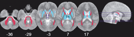
The figure above shows the brain structures affected by increased free water content in obesity overlayed on a mean quantitative proton density map (A, anterior limb of internal capsule; D, dorsal striatum, including caudate and putamen; F, fornix; H, lateral hypothalamus; MCP, middle cerebellar peduncle; P, posterior thalamic radiation; Th, thalamus).
Original publication:
Investigating obesity-associated brain inflammation using quantitative water content mapping