JUQEBOX (Juelicher Quantitative ToolBox)
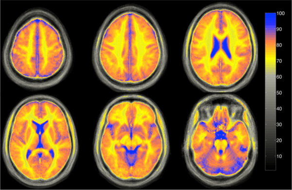
JUQEBOX (Juelicher Quantitative ToolBox) employs recent advances in quantitative MRI (qMRI) in order to provide high quality maps of tissue specific parameters (T1,T2*) as well as free water content distribution (often abbreviated for simplicity as proton density - PD). Different processing algorithms are established on the basis of well-defined qMRI protocols such as 2D/3D GRE (Abbas 2014-2015, Oros 2013) or QUTE-TAPIR (Shah 2001-2008).
The specialist software is capable of handling data from high-fields (>1.5T), where dependence between transmitter voltage and received signal is no longer obvious. For this purpose, transmit field inhomogeneity (B1+) as well as receiver inhomogeneity (B1-) and residual non-uniformity must be corrected (Gras 2013, Abbas 2014). These corrections can be applied, not only as a part of PD/T1/T2* mapping procedure, but also as an independent protocol which broadens the scope of applications for the toolbox.
JUQEBOX can be set-up to use computing clusters, in particular GPGPU devices compatible with OpenCL (virtually every device). OpenCL kernels (executed on valid hardware) can speed up the critical, most exhausting elements of the processing chain by more than 100 times.
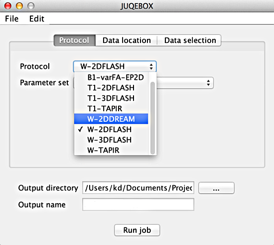
Core functionalities of JUQEBOX were developed in Matlab and front-end (GUI) in Java. This, together with good documentation, makes JuqeBox very easy to learn for new users.
The final outcome of JUQEBOX - calculated quantitative maps (PD,T1,T2*) are perfect input for further systematic image analysis (e.g. statistical analyses, construction of brain atlases, etc.). Tissue specific parameters maps (T1,T2*,PD) can be very useful in clinical applications such as detection of cerebral oedema in brain diseases or abnormal water content analysis in patient cohorts.
REFERENCES
- N. J. Shah, H. Neeb, M. Zaitsev, S. Steinhoff, G. Kircheis, K. Amunts, D. Häussinger, and K. Zilles, “Quantitative T1 mapping of hepatic encephalopathy using magnetic resonance imaging.,” Hepatology (Baltimore, Md.), vol. 38, no. 5, pp. 1219–1226, Nov. 2003.
- H. Neeb, K. Zilles, and N. J. Shah, “A new method for fast quantitative mapping of absolute water content in vivo.,” NeuroImage, vol. 31, no. 3, pp. 1156–68, Jul. 2006.
- V. Gras et al., “Quantitative water content mapping at 1.5 & 3 Tesla field strength,” ISMRM, no. n Proceedings of the 19th Annual Meeting of Montreal, Canada, 2011. p. 4449, 2011.
- Z. Abbas, V. Gras, K. Möllenhoff, F. Keil, A.-M. Oros-Peusquens, and N. J. Shah, “Analysis of proton-density bias corrections based on T1 measurement for robust quantification of water content in the brain at 3 Tesla.,” Magnetic resonance in medicine : official journal of the Society of Magnetic Resonance in Medicine / Society of Magnetic Resonance in Medicine, vol. 00, no. 6, pp. 1735–45, Jan. 2014.
- Z. Abbas, V. Gras, K. Möllenhoff, A.-M. Oros-Peusquens, and N. J. Shah, “Quantitative water content mapping at clinically relevant field strengths: A comparative study at 1.5T and 3T.,” NeuroImage, vol. 106, pp. 404–13, Feb. 2015.
- V. Gras, Z. Abbas, and N. J. Shah, “Spoiled FLASH MRI with Slice Selective Excitation : Signal Equation with a Correction Term,” Concepts in Magnetic Resonance, vol. 42, no. April, pp. 89–100, 2013.
- R. Venkatesan, W. Lin, and E. M. Haacke, “Accurate determination of spin-density and T1 in the presence of RF-field inhomogeneities and flip-angle miscalibration.,” Magnetic resonance in medicine : official journal of the Society of Magnetic Resonance in Medicine / Society of Magnetic Resonance in Medicine, vol. 40, no. 4, pp. 592–602, Oct. 1998.
- H. Neeb, V. Ermer, T. Stocker, and N. J. J. J. Shah, “Fast quantitative mapping of absolute water content with full brain coverage,” NeuroImage, vol. 42, no. 3, pp. 1094–1109, Sep. 2008.
- A. Oros-Peusquens, F. Keil, K. J. Langen, H. Herzog, G. Stoffels, C. Weiss, and N. J. Shah, “Fast and accurate water content and T2* mapping in brain tumours localised with FET-PET,” Nuclear Instruments and Methods in Physics Research Section A: Accelerators, Spectrometers, Detectors and Associated Equipment, pp. 1–6, Oct. 2013.