Quantitative cerebral water content mapping
Information relating to water content in the brain is potentially present in any proton MR image due to the fact the magnetisation, M0, induced by the main magnetic field is proportional to the proton density (PD).
However, in practice, estimating M0 necessitates a number of corrections to suppress the influence of the relaxation times T1 and T2 and to ensure that static field or RF field inhomogeneity does not bias the estimation.
Here at the INM-4, researchers are working on two sequences to investigate water content mapping, namely the TAPIR sequence, originally created to produce maps of the T1 relaxation time, and the QUTE sequence, designed to estimated the T2* relaxation time.
The QUTE sequence, is also known today as mutli-echo gradient echo (GRE) and is provided by the manufacturer of the scanner.
The two approaches give rise to four water content protocols
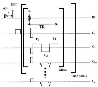
The TAPIR water content mapping protocol
The M0-weighted 2D-GRE protocol
The T1-weigthed 2D-GRE protocol
The 3D-GRE protocol
The TAPIR, the M0-w. 2D-GRE and the T1-w. 2D-GRE protocols provide water content maps with an in-plane resolution of 1 mm and a slice thickness of 2 mm whereas the 3D-GRE protocol was verified to up to 1 mm isotropic resolution. However, the 3D-GRE protocol does not yet offer the same accuracy as the 2D-protocols. The TAPIR protocol is well suited for high in-plane resolution (e.g., 0.7 mm) when partial brain coverage is acceptable. The 2D-GRE protocol offers the best alternative for rapid whole brain water content imaging.
It is anticipate that the development of these sequences will help researchers obtain brain water maps with unprecedented resolution, helping to increase our understanding of water in the brain and to aid accurate diagnosis and treatment planning of pathologies associated with changes in brain water content.