fMRI & EEG
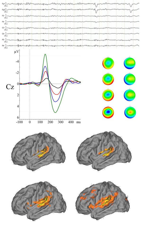
The main aim of simultaneous electroencephalogram (EEG) and functional magnetic resonance imaging (fMRI) recording is to combine the high temporal resolution of EEG and the spatial resolution of fMRI. EEG has a temporal resolution in the order of ms, whereas the temporal resolution of the fMRI blood oxygenation level dependent (BOLD) response is in the order of seconds. However, the spatial resolution of EEG is poor in comparison to fMRI. By using these imaging techniques in combination, we are able to investigate both, the time course and exact location of brain activity in response to a stimulus. This way, the simultaneous recording of electrophysiological and hemodynamic responses to the same neural event affords maximal exploitation of the complementary strengths of EEG and fMRI. In addition, behavioural responses such as reaction time or accuracy are also recorded, thus providing a tri-modal representation of subjects' responses to any given event in an experiment. This provides a rich source of data for exploring cognitive function. Recent work in the group has focused on investigating analysis techniques for successful combination of the two data sets. Ongoing and future work focuses on applying these analysis techniques to other research areas, such as the effects of pharmacological challenges on cognition and the investigation of different patient groups.