EPIK and high-resolution fMRI
Demonstration of EPIK in fMRI
EPI is in widespread use in MR studies acquiring time series data due to its capability of achieving a relatively high temporal resolution. As an approach to improve its performance, EPIK (EPI with Keyhole) was proposed by Shah et al. [1,2] and validated by Zaitsev et al. at 1.5T [3,4]. In EPIK acquisition, each measurement scans the central k-space region completely, whilst the peripheral k-space regions are sparsely sampled resembling a multi-shot scheme. A complete k-space can be reconstructed by sharing the sparse region data from the consecutive scans; importantly, use of a sliding window ensures that only the last few shots contribute to any given image. With this strategy, we can achieve a higher temporal resolution, a reduced signal-loss and reduced image artefacts compared to the single-shot EPI.
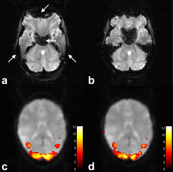
As shown in the Figs. 1a and b, the EPIK image had reduced geometric distortions than the EPI image, particularly around the regions marked by white arrows. The feasibility of using EPIK for fMRI has been demonstrated with a visual checkerboard paradigm. Figures 1c and d suggest that visually-induced brain activations were consistently detected from both imaging methods and the result of EPIK has features very comparable to that of EPI [5].
High-resolution fMRI using accelerated EPIK
The EPIK acquisition can be further accelerated by parallel imaging and partial Fourier techniques to achieve even higher resolution. By pushing the limits to the highest possible resolution with the acceleration techniques, accelerated EPIK achieved 1 x 1 mm in-plane resolution whilst accelerated EPI achieved 1.25 x 1.25 mm in-plane resolution. Furthermore, when the same TR (3s) was given, more number of slices were obtained with accelerated EPIK (32) than with accelerated EPI (28).
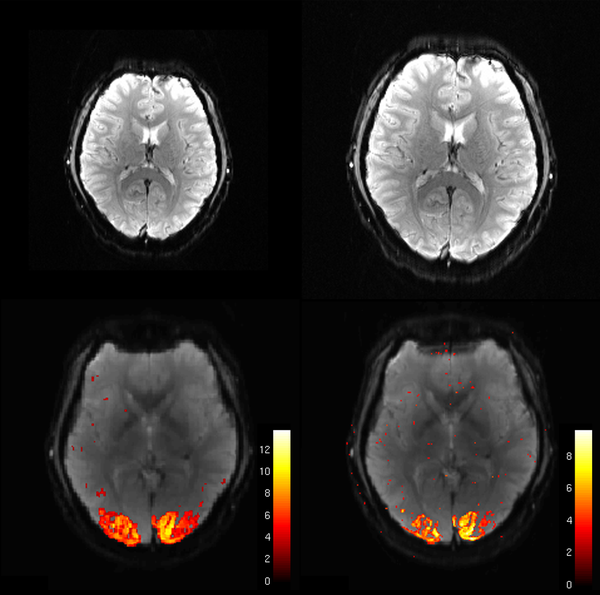
As shown in the Figs 2a and b, the image from each imaging method was reconstructed without any severe distortions however, the anatomical features are more clearly represented in the accelerated EPIK image than in the accelerated EPI image. The visual inspection of the functional maps (Figs. 2c and d) suggests that the result from accelerated EPIK depicts more precise mapping of functional quantities along with the cortices regions than that from accelerated EPI.
In this project, we will implement not only single-contrast EPIK, but also dual-contrast and multi-contrast EPIK sequences. By using these sequences, we can obtain fMRI images with various contrasts and perform quantitative analyses.
References
- Shah NJ, Zilles K. Verfahren zur Untersuchung eines Objektes mittels Erfassung des Ortsfrequenzraumes. German Patent Application 2003;No. 199 62 845 C2. Shah NJ, Zilles K. Verfahren zur Untersuchung eines Objektes mittels Erfassung des Ortsfrequenzraumes. German Patent Application 2003;No. 199 62 845 C2.
- Shah NJ, Zilles K. Imaging process in the spatial frequency space and useful for examining the properties of object. USA Patent Application 2004;No. 6781372 B2.
- Zaitsev M, Zilles K, Shah NJ. Shared k-space echo planar imaging with keyhole. Mag. Reson. Med. 2001;45:109-117.
- Zaitsev M, Arcy JD, Collins DJ, Leach MO, Zilles K, Shah NJ. Dual-contrast echo planar imaging with keyhole: application to dynamic contrast-enhanced perfusion studies. Phys. Med. Biol. 2005;50:4491-4505.
- Yun S, Reske M, Vahedipour K, Warbrick T, Shah NJ. Parallel imaging acceleration of EPIK for reduced image distortions in fMRI. NeuroImage 2013;73;135-143.