Research Videos
Emergence of Metachronal Waves in Cilia Arrays

Emergence of Metachronal Waves in Cilia Arrays Using Large-Scale Hydrodynamic Simulations
Related publication: PNAS 110 (12) 4470-4475 (2013)
Visualization of tissue competition

Two tissues that only differ by their homeostatic pressure compete for space. The blue tissue has the higher homeostatic pressure than the red, and thus grows towards the right. The field of view advances with the tissue interface (the grey squares moving towards the left represent the non-moving substrate).
Related publication
N.J.Phys. 18:83020 (2016)
Interface dynamics of competing tissues
Particle based tissue growth model
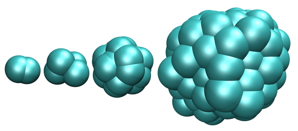
Cells are represented by point particles that repel themselves in order to grow. When a size threshold is reached, the cell divides. Apoptosis is implemented as a constant rate of cell removal. Otherwise cells behave like sticky soft colloids.
Related publication
Phys. Biol. 8:26014 (2011)
Dissipative particle dynamics simulations for biological tissues: rheology and competition
Tensile homeostatic state

Due to the free interface, cell division is favored close to the surface. If division rate in the bulk is smaler than the apoptosis rate this leads to a steady state. If instead the surface is removed, by attaching to a no-slip surface, the tissue shrinks, pullin in the piston, and reaching a tensile steady state.
Related Publication
Europhys. Lett. 109, 58005 (2015)
Tissue homeostasis: A tensile state
Ciliated Microswimmer
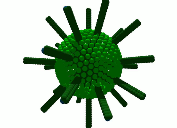
Cilia, grafted on a spherical surface, propell the swimmer foreward by their beating motion.
Tensile growth of a tissue colony
Cells grow by devision, but in combination with motility forces, the growing colony can be under tension.
Related Publication:
PROCEEDINGS OF THE NATIONAL ACADEMY OF SCIENCESVol. 110 | No. 7 (2013)
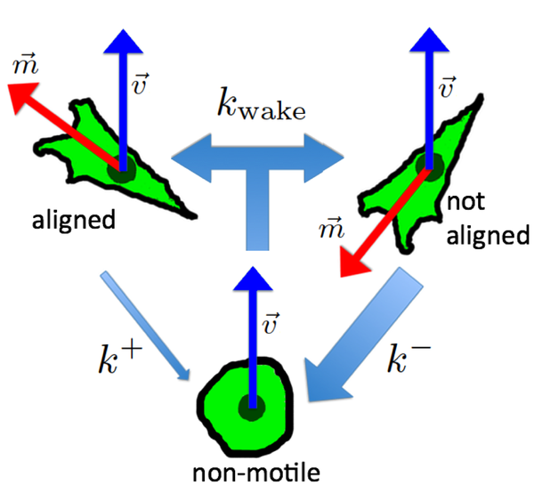
Alignment of cellular motility forces with tissue flow as a mechanism for efficient wound healing